I AM
JOÃO VITOR M. FERNANDEZ
The constantly evolving field of technology requires a combination of hard and soft skills. Here are mine:
Artificial intelligence skills
Data analysis skills
Radiation Oncology: TPS and R&V
Radiation Oncology: IGRT Technology Experience
Radiation Oncology: Machine QA, PSQA and dosimetry skills
Worked as a clinical medical physicist / deputy radiation safty officer (RSO) at Rota dos Bandeirantes Regional Hospital. As Deputy RSO, I advise the License Holder, the Medical Technical Responsible, and the principal RPO on all matters related to safety and radiation protection within the Radiotherapy Department.
Worked as a medical physicist in radiotherapy in Elekta environment, doing conformal and VMAT treatment planning (using Monaco v5.11.03). Prepared treatment records and performed quality assurance, including periodic dosimetry (following IAEA TRS-398). Developed strong decision-making abilities, flexibility in adapting to challenging clinical environments, and excellent organizational skills while managing a high patient workload in a resource-limited setting. Gained solid experience in troubleshooting critical mechanical and dosimetric issues on the Elekta platform.
Shadowing experience in a clinical environment that provides care to cancer patients in Brazil’s public health system (SUS). Learned about 2D radiotherapy treatment planning for breast cancer using CAT3D software and 3D conformal planning with the Eclipse treatment planning system (Varian). Also participated in starting treatments, performing machine adjustments (Clinac iX, 600C, and Unique), and doing dosimetry following IAEA TRS-398 guidelines.
Extracurricular internship at Vila Nova Star Hospital (São Paulo, Brazil), where I learned about radiotherapy treatments on three different platforms: TrueBeam (Varian), TomoTherapy, and CyberKnife (Accuray). Observed treatment planning using Eclipse and Precision, and helped with quality assurance on each machine. Main highlights were a liver SBRT case with tracking on CyberKnife and a neuroaxis treatment done with TomoTherapy.
Extracurricular internship at Sievert Laboratory and Environmental Services (São Paulo, Brazil), focused on learning more about radiation dose estimation for imaging and environmental exposure. Worked on collecting data, running simulations, and calculating radiation doses from inhalation of naturally occurring radioactive materials (NORMs) by miners. Also did a comparison of different computational methods used for dose calculation in this area.
Mandatory curricular internship divided into three areas of medical physics: Radiotherapy (120 hours), Diagnostic Radiology (120 hours), and Nuclear Medicine (120 hours). Activities included following the daily work in each department and doing specific quality control tests. In the radiotherapy part, I also worked on conformal treatment planning for prostate and breast cancer using XiO software.
Part of the mandatory curricular internship in radiotherapy was done at CAISM. Activities included observing quality control routines, dosimetry procedures, and doing 2D and 3D conformal treatment planning for breast cancer using XiO software (Elekta).
Observed the daily work of the radiotherapy department, taking part in all steps of the treatment process, including simulation and treatment planning for external beam radiotherapy using 3DCRT, IMRT, and VMAT, as well as HDR brachytherapy. Also learned about special treatments like Total Body Irradiation (TBI) and blood irradiation for transplants. This experience was a strong motivation for me to follow a career in radiotherapy.
Postgraduate lato sensu program with 362 hours at Data Science Academy (DSA), focused on improving the use of artificial intelligence (AI) in healthcare, especially radiotherapy workflows and expanding my knowledge beyond my main field. The course covered topics such as advanced AI methods, Deep Learning with Python and C++, medical image analysis, large language models (LLMs), creating and using predictive models, ethical issues, data privacy in AI, and practical projects with real healthcare data.
Completed 5,760 hours of specialized training in a modern clinical Varian environment. Gained practical experience in simulation, treatment planning, and dose delivery. Worked with different treatments such as SABR, SRS, and also with brachytherapy (LDR and HDR). Developed skills in IGRT technologies like CBCT, ExacTrac, and SGRT using OSMS. Performed monthly quality control based on TG-198, dosimetry following TRS-398, and patient-specific quality assurance (PSQA). My final project was about validating the 3D printing process of customized bolus using PLA, it was presented in 29º Brazilian Congress of Medical Physics
A 300-hour professional development course at Data Science Academy (DSA) aimed at learning how to integrate artificial intelligence (AI) algorithms into radiotherapy, covering treatment planning, risk management, and quality control. The curriculum included health data analysis, applying machine learning to data, medical image analysis with AI, and some automation in medicine using AI and RPA of clinical workflows and processes. I was invited to participate in the DSA podcast to share my insights on artificial intelligence in the field of radiotherapy.
Bachelor’s degree in Physics with a minor in Medical Physics plus 930 hours of extracurricular courses including Modern Optics and Photonics, Quantum Technology, Computer-Aided Technical Drawing, Laser Physics, Electronics, among others. During my undergraduate studies, I actively participated in the research project Fabrication of interdigital transducers and growth of ZnO films for the generation of surface acoustic waves Additionally, I developed an automated temperature-controlled TLD irradiator which supported my undergraduate research focused on a study of the thermoluminescent properties of apatite from the Durango region in Mexico.
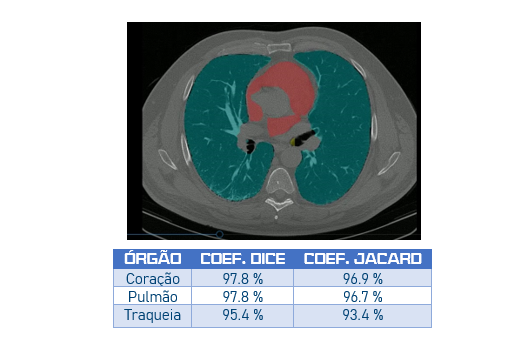
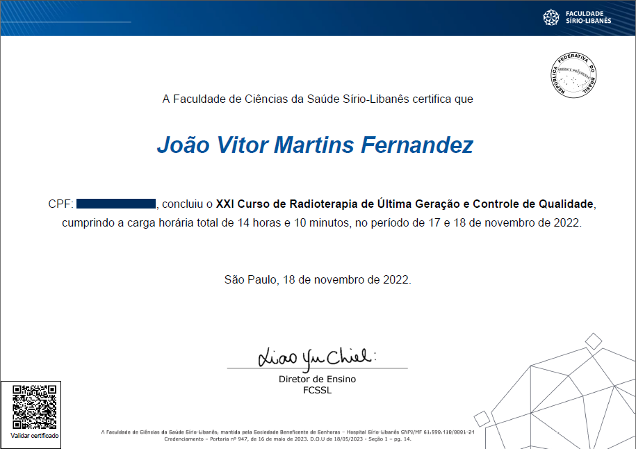
This traditional annual course hosted by Hospital Sírio-Libanês provides updates and fosters discussion on cutting-edge advances in radiation therapy and associated quality control protocols.
Covered key pediatric tumor types (e.g. medulloblastoma), treatment planning strategies, team emotional preparedness, and patient-centered care approaches, including imobilization mask decoration to support a more humanized experience.
Focused on the entire treatment workflow — from simulation/immobilization to quality assurance — this course also included hands-on planning cases using the Shulman method to achieve optimal treatment plans.
Continuing education course on radiosurgery techniques, covering technologies employed, treatment planning approaches, quality assurance procedures, and clinical cases of tumors in the central nervous system, as well as functional radiosurgery (e.g., depression, trigeminal neuralgia, and Parkinson's disease).
This workshop addressed the theoretical and practical aspects of low-dose-rate (LDR) brachytherapy for prostate cancer. In the 2023 edition, I had the honor of participating as a resident, contributing to volume analysis, needle preparation, and assisting in the implantation procedure.
This 40 hour course offered by the Brazilian National Cancer Institute (INCA) aimed to develop competencies in the clinical application of electron beams in radiation therapy. The curriculum covered the fundamental physics of electrons, clinical indications, and dosimetry protocols.
Radiological emergencies, such as the loss of a brachytherapy source, must be rigorously prevented. Nevertheless, in the event they do occur, they demand prompt and effective response measures. This course offered by the IRD addressed the entire response framework — from preparedness and planning to the management of emergency situations.
With advances in imaging technologies, it has become possible to diagnose tumors with greater precision and to treat them using specialized techniques such as stereotactic radiosurgery (SRS). This short course introduced the Elements software, Brainlab's solution for SRS planning.
To uphold Hippocrates' maxim "Primum non nocere" ("First, do no harm"), high-quality radiotherapy must be delivered safely. This course addressed several tools for mitigating incidents, such as FMEA (Failure Modes and Effects Analysis) and FTA (Fault Tree Analysis), as well as incident learning systems in radiotherapy, such as SAFRON.
Since radiotherapy relies primarily on CT imaging, it is essential to understand the parameters that affect image quality, as well as dose optimization techniques and specific protocols — particularly for pediatric patients and pregnant individuals.
This hands-on workshop, offered by Varian during the XXV Brazilian Congress of Medical Physics (CBFM), covered radiotherapy treatment planning using Varian’s state-of-the-art technology. Topics included automatic contouring and AI-assisted treatment planning.
This course reviewed core radiobiology concepts and highlighted current advances in the field, including the radiobiological foundations of emerging modalities such as FLASH and LATTICE radiotherapy.
This course covered the basic principles of radiotherapy, indications, patient selection, curative and palliative treatment, emergencies and benign diseases, and radiotherapy techniques: 3DCRT, IMRT, VMAT, SBRT, and SABR.
